Physical examination
Aberrant neurological behaviour can occur as a result of metastases to nervous tissue.
Lameness may occur as a result of hypertrophic osteopathy in which new bone forms at a site remote from the primary tumour. The limping may improve after the lung tumour is removed.
Often a lung tumour does not cause abnormalities on auscultation (examination with the stethoscope) unless the process is extensive and / or because of fluid build-up in the lungs or the thoracic cavity.
Blood analysis
Often no abnormalities are found on blood tests.
Imaging
Radiography
The majority of lung tumours are diagnosed by chest radiographs. In a study involving more than 210 dogs with a primary lung tumour, more than half (53.8%) were found in a single lung lobe and 37.1% in multiple lobes.
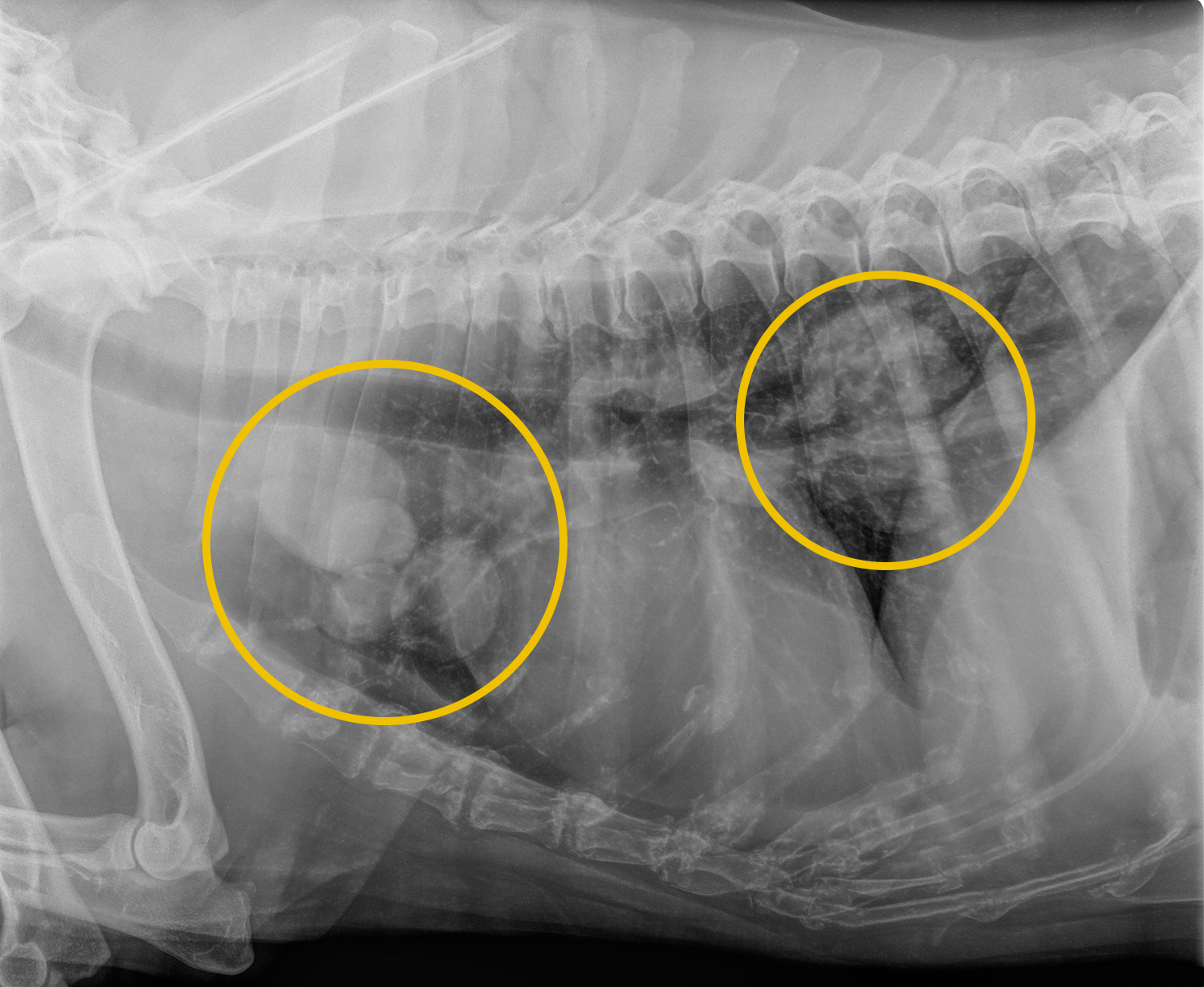
The round white spots on the radiography indicate the presence of metastases
Ultrasound
Chest ultrasound can be used in some cases to assess a lung tumour or to obtain a tissue sample or aspirate.
CT-scan
A CT scan is recommended prior to surgery to be able to draw up a precise treatment plan. After all, CT scans can detect lumps in the lungs with dimensions of about 1 mm, while with radiographs this can only be done starting from 7-9 mm. Furthermore, CT scans of the chest are more accurate than chest radiographs in detecting metastases to nearby lymph nodes.
Sampling
If fluid accumulation in the lungs is present, a sample should be taken here.
Furthermore, broncho-alveolar lavages (the administration of fluid through a tube to the lungs, after which this fluid is withdrawn) could help to diagnose lung tumours. If a lung tumour is present, there is a good chance that the accumulated fluid contains cancer cells that can subsequently be recognized under a microscope. However, this approach is based on results obtained from a small group of dogs (14).
A fine needle aspirate of the lung mass provides a diagnosis in 38-90% of cases but is not possible in all cases due to the location of the presumed tumour.
A lung tissue sample prior to surgery can be done to find out if the nodule is a lung metastasis or a primary tumour. In the case of metastasis, surgery comprising of the removal of a part of the lungs is generally disregarded as an option.
The sampling itself can be done via a biopsy needle, an endoscope in the airways or an operation during which a part of the lung is removed. Tissue sampling of the lymph nodes at the branch between both lungs is recommended because spread to the lymph nodes significantly affects the prognosis and usually performed during surgery.
Histology
It is not easy to distinguish between a tumour that originates in the lungs (primary tumour) and a spread of another type of tumour, because a tissue sample is required for this. By means of a tissue staining, antibodies can be used that can distinguish between a primary lung tumour and a metastasis.