During Magnetic Resonance Imaging (MRI) the patient is placed in a magnetic field and via the radio waves a 3D image is created based on the water molecules in the patient. During the scan perpendicular or longitudinal slices of an organ or body area are made via radio waves. This imaging technique shows how elaborate the tumour is and whether it grows into the surrounding tissues. An MRI scan is capable of showing a more detailed representation of a certain part of the body than ultrasound or radiography, especially for soft tissues and the brain. An intravenous contrast fluid can also be administered to the patient to show certain anomalies on the MRI scan even more clearly.
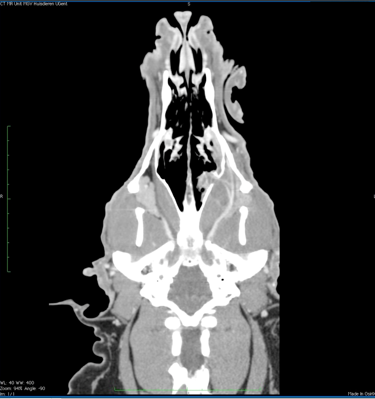
MRI of a dog’s head